Phylum Porifera:
 |
| Commercial sponge fibers at 25x. |
Sponges are made up of only four simple, interdependent cells that work together. Going from the inside of the sponge to the outside, the first cells are collar cells that line the canals inside the sponge where flagella help to pump through water. The second cells are porocytes, they make up the pores of the sponge that allow for the water to enter. Next are epidermal cells that form the outer skin. Lastly, there are
ameobocytes between the epidermal and collar cells which carry out the functions of the sponge and form spicules which are the sponge's skeletal fibers.
 |
| Dried sponge fibers at 25x. |
The two imbedded images of sponges are very similar in nature. You can see the fibers which are dyed blue in the first picture and distinct in the dried sponge fibers. The fibers are made up by the four interdependent cells talking about above. The pores that bring it water are also visible in both views shown by the gaps in fibers. Back in the day, sponges were plucked out of their habitat to use as our everyday kitchen sponges until we were able to synthesize replicas of their fibers to create the commercial sponge shown above.
Three different species of sponges:
The Row Pore Rope Sponge, scientifically know as,
Aplysina cauliformis is from the class
Demospongiae. It grows from four to eight feet tall with long, branching rope-like tips at depths from 40 to 130 feet. This sponge is stiff and hard with varying colors from reds to purples. They are mainly found off the
coast of Florida to islands in the Caribbean Sea.
The yellow tube sponge, or sulphur sponge, scientifically known as
Aplysina fistularis, is a large sessile sponge that can grow to over three feet tall. It can reproduce both sexually and asexually. If a piece is broken off, it can reattach to a new part of the reef and start growing. This sponge
thrives at depths less than 100 feet because the stronger currents of the waves helps to bring more water in.
Giant barrel sponge, or the
Xestospongia muta, can grow up to six feet. It is shaped like a bowl providing a habitat for many different species. It is also
hermaphroditic, meaning that each sponge produces both sperm and eggs. They release their own sperm into the water keeping the eggs within. Their eggs get fertilized through filter feeding, they filter the sperm from others our and use to to fertilize their own eggs that they keep within. Once the eggs are mature they get released so they can settle elsewhere and create new sponges. These sponges are known to lives up to 2000 years, but maybe even longer.
Some commonalities that all these sponges have are that they are found mainly around the Caribbean expanding upwards all the way to Florida. They all feed through filter feeding where they bring the ocean water in through their pores to obtain nutrients and oxygen by filtering it out from the water.
Sponges do not have a nervous, digestive, or circulatory system, instead they filter feed. They have ostia, tiny pores, in their outermost wall that bring in water, sponges need a constant flow of water to get food and oxygen. It also is a way to remove waste out of their bodies. The cells in the wall filter the food out from the water by flagella pushing it through the gaps bringing it to the osculum (mouth). Sponges get their coloring depending on the algae around them. When they are greenish it is because of a symbiotic relationship with green algae within the sponge. The purple and pinkish colors come from symbiotic blue-green algae. This is why sponges are not found at deeper depths of water so that there is sunlight to allow for the photosynthesis of the algae.
Phylum Cnidaria:
Cnidarians come in two forms, polyp or medusa which is essentially an upside down polyp. The difference is that the polyp form is attached to a surface while the medusa is free-floating. The
Portuguese man o' war is an example where both medusa and polyp stages are present in a single colony.
 |
| Budding hydra at 4 x 0.10 magnification. |
Hydra are freshwater invertebrates in the class Hydrozoa, phylum Cnidaria. Their bodies are a hollow cylinder consisting of the digestive tract with a mouth at the top with surrounding tentacles that bring in the food into mouth. At the bottom of cylinder is a
basal disc which secretes a sticky substance that allows for the hydra to attach to a hard substance. They are not always attached to a surface and can move around by gliding or un-attaching allowing for them to float around before reattaching somewhere else. Shown to the side is an image taken by myself of a budding hydra. This is how they
reproduce, the budding forms a mouth and tentacles where it will then cut off of at the base, forming a new individual.
 |
| Obelia at 10x/0.25 magnification. |
 |
| Obelia at 4x magnification. |
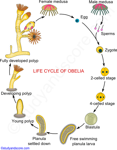
Obelia is a colonial
coelenterate that lives in both fresh and salt water habitats. Its structure is branching stems composing of
minute cups where the polyps sit in. Obelia has a medusa form and is both sessile and mobile. The main difference between Obelia and Hydra is that Hydra's main body form is the polyp but Obelia have both polyps and medusa in its life cycle.
 |
| Upside down mangrove jelly moving. |
The upside down mangrove jelly,
Cassiopea xamachana, is a Cnidarian in the class
Scyphozoa. The highest density of their
geographical distribution is in the Caribbean but they have been expanding to other tropical waters. They got their name from using the muddy
substrata on mangrove leaves to rest upon. Their coloration helps them to camouflage as their upside down medusa form resembles vegetation or algae under the water. These jellyfish also have cnidocytes, venomous stinging cells. A single sting from one is not deadly but, when one is triggered to sting, a swarm of them come up posing a great danger
 |
| A sea anemones polyp form at 4x. |
 |
| A close up view of the tentacles. |
Sea anemones,
Actiniaria, are from the class
Anthozoa and take the polyp form of Cnidarians. Anemones have a cylindrical body with a pedal disk that they use to attach to a hard base and
nematocysts in their tentacles to paralyze and capture their meals. They thrive in the warmer waters where they can be found in the most numerous amounts being larger and brighter than others.
 |
| Hydroid colony at 25x. |
Hydroids are invertebrates from the class
Hydrozoa, they are a
colonial organism. These colonies can secrete calcium carbonate skeletons for some added protection and some are so integrated with one another that they act like a single organism. They have
three different stages of life: a free swimming larva only about a millimeter long that settles and metamorphoses into a sessile, usually colonial, polyp stage, which then turns into a gamete-producing male or female medusa.
 |
| Hard coral structure at 25x magnification. |
The skeletons of coral reefs are comprised of thin layers of calcium carbonate. The polyps form a "
living mat" over this skeleton. Most corals have learn bodies, they get their coloration from
zooxanthellae, tiny algae, that live inside their tissues. They have a
mutualistic relationship with one another, the coral provides an environment for the algae and the compounds they need to be able to photosynthesize while the algae produce oxygen and help the corals remove wastes.


.jpg)

.jpg#/media/File:Aplysina_fistularis_(Yellow_Tube_Sponge).jpg)

.jpg)

_(San_Salvador_Island,_Bahamas)_4_(15896072120).jpg#/media/File:Physalia_phyalis_(Portuguese_man-of-war)_(San_Salvador_Island,_Bahamas)_4_(15896072120).jpg)


Comments
Post a Comment